Endometriosis: Symptoms, Diagnosis, Treatment & Fertility
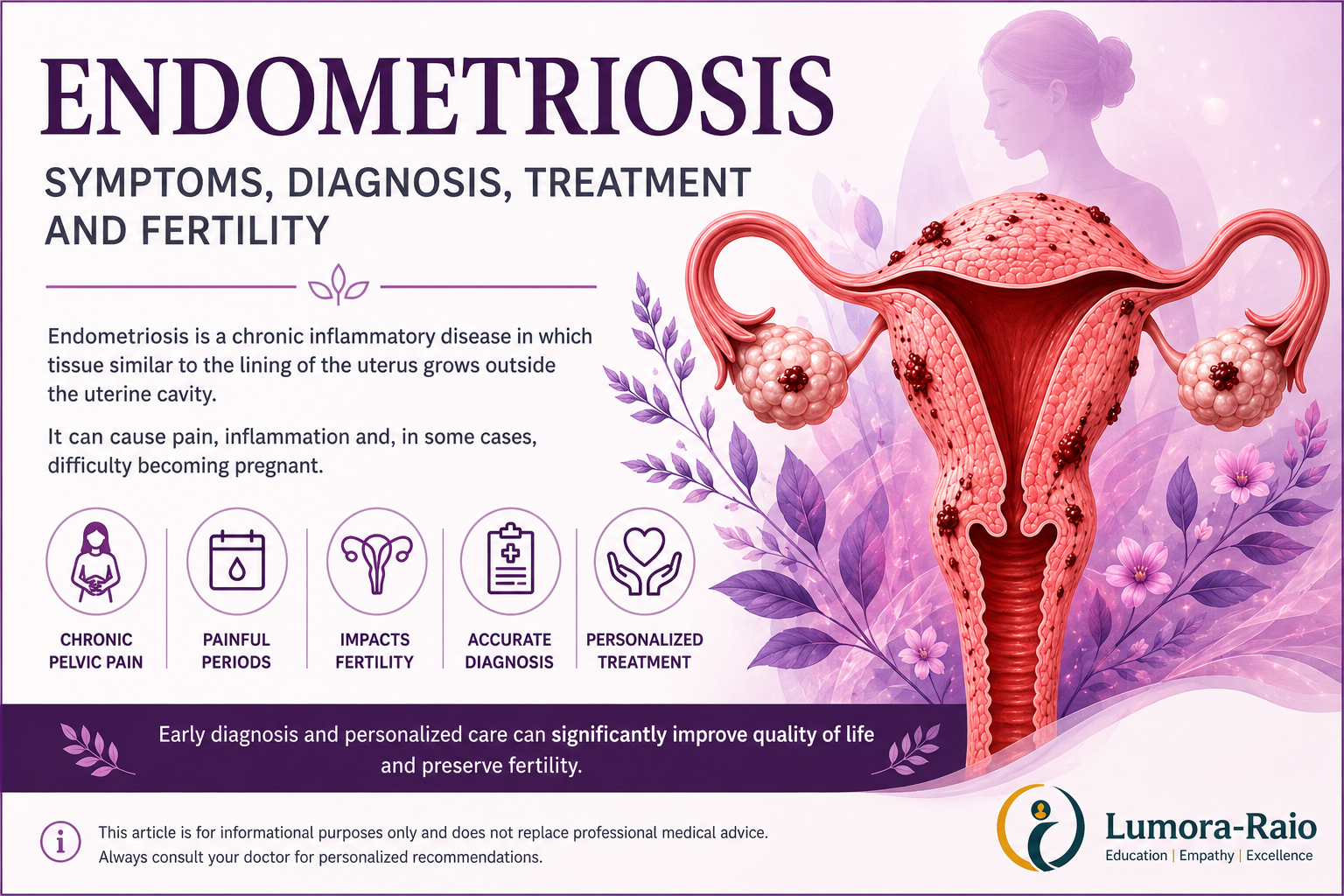
Endometriosis is a chronic inflammatory gynaecological disease characterised by the presence of endometrial-like tissue outside the uterus. Unlike the endometrium, which normally lines the uterine cavity, endometriosis lesions may develop on the pelvic peritoneum, ovaries, uterosacral ligaments, fallopian tubes, bladder, bowel and, more rarely, outside the pelvis, including the diaphragm and chest. [1–4]
The disease may cause inflammation, fibrosis, adhesions, ovarian endometriomas, chronic pain and fertility problems. Symptoms and their impact vary greatly between individuals.
The World Health Organization estimates that endometriosis affects approximately 10% of women of reproductive age worldwide—around 190 million people. Symptoms may begin during adolescence, sometimes soon after the first menstrual period, and may persist after menopause. Endometriosis can also affect transgender men and non-binary people who menstruate. [1]
Endometriosis is not simply a menstrual disorder. It can affect physical health, sexual life, fertility, emotional wellbeing, relationships, education and employment.
What types of endometriosis are there?
Endometriosis is commonly described according to the location and appearance of the lesions:
Superficial peritoneal endometriosis
Small lesions develop on the surface of the pelvic peritoneum or pelvic organs.
Ovarian endometriosis
Endometriosis affects one or both ovaries and may form cysts called endometriomas.
Deep endometriosis
Lesions extend beneath the surface of the peritoneum and may involve the uterosacral ligaments, vagina, bowel, bladder, ureters or pelvic nerves.
Extrapelvic endometriosis
Rarely, lesions occur outside the pelvis, including on the diaphragm, abdominal wall or in the chest.
The anatomical extent of the disease does not reliably predict the intensity of pain or its effect on quality of life.
What are the symptoms of endometriosis?
Symptoms differ considerably from one person to another. Some people have extensive endometriosis with few symptoms, while others experience severe pain despite apparently limited disease.
Common symptoms include:
- severe or progressively worsening menstrual pain;
- chronic pelvic pain;
- lower abdominal or lower back pain;
- pain during or after sexual intercourse;
- painful bowel movements, particularly during menstruation;
- cyclical bloating, diarrhoea, constipation or nausea;
- pain during urination, especially during menstruation;
- cyclical blood in the urine or rectal bleeding;
- heavy menstrual bleeding or spotting;
- fatigue;
- difficulty becoming pregnant;
- reduced quality of life;
- anxiety, depression or emotional distress associated with chronic pain and delayed diagnosis.
Symptoms involving the bowel or bladder may resemble gastrointestinal or urinary disorders. Endometriosis can therefore coexist with, or initially be mistaken for, conditions such as irritable bowel syndrome, painful bladder syndrome, pelvic inflammatory disease, adenomyosis or musculoskeletal pelvic pain.
The intensity of pain does not necessarily correspond to the number, size or location of the lesions.
Endometriosis in adolescents
Endometriosis can occur in adolescents and should be considered when menstrual pain:
- interferes with school, sport, sleep or normal activities;
- does not improve adequately with appropriate analgesic or hormonal treatment;
- is accompanied by chronic or cyclical pelvic, bowel or urinary symptoms;
- becomes progressively more severe;
- is associated with a family history of endometriosis.
Severe menstrual pain should not automatically be dismissed as a normal part of adolescence.
Current ACOG guidance applies clinical and imaging-based diagnostic principles to adolescents as well as adults, while recognising that the available evidence in younger patients remains more limited.
Who is more likely to develop endometriosis?
Endometriosis can affect anyone born with female reproductive organs, regardless of ethnicity or social background.
Factors associated with a higher likelihood of endometriosis include:
- a first-degree relative with endometriosis;
- early onset of menstruation;
- shorter menstrual cycles;
- prolonged or heavy menstrual bleeding;
- fewer or no previous pregnancies;
- anatomical conditions that obstruct menstrual flow.
A family history is particularly relevant. Having an affected first-degree relative increases the likelihood of developing the condition.
These factors are associations, not proof that a person will develop endometriosis. Many affected individuals have no identifiable risk factor.
What causes endometriosis?
The exact cause remains unknown. No single theory explains every form of the disease.
Several biological mechanisms may contribute:
- retrograde menstruation, in which menstrual fluid travels through the fallopian tubes into the pelvic cavity;
- genetic susceptibility;
- altered immune and inflammatory responses;
- hormonal influences, particularly oestrogen-dependent activity;
- transformation of peritoneal or embryonic cells into endometrial-like cells;
- spread of endometrial-like cells through blood or lymphatic vessels;
- persistence of lesions through abnormal inflammation, nerve formation and blood-vessel development.
Endometriosis is therefore considered a multifactorial disease involving genetic, hormonal, immune, inflammatory and environmental mechanisms. Research is continuing, and none of the proposed mechanisms alone has been proven to account for all cases.
Can endometriosis cause infertility?
Yes.
Although many women with endometriosis conceive naturally, the disease is associated with reduced fertility.
Reduced fertility may result from several mechanisms, including:
- pelvic inflammation;
- adhesions and distortion of pelvic anatomy;
- fallopian tube dysfunction;
- ovarian endometriomas;
- reduced ovarian reserve;
- impaired ovulation or reduced oocyte quality;
- altered interaction between sperm and oocyte;
- impaired embryo implantation.
Endometriosis is reported in approximately 25–50% of women evaluated for infertility, although prevalence varies according to the population studied and the diagnostic method used. This does not mean that endometriosis is always the sole cause of infertility.
Fertility treatment
Management depends on:
- age;
- duration of infertility;
- ovarian reserve;
- location and extent of endometriosis;
- fallopian-tube function;
- semen analysis;
- previous surgery;
- pain symptoms;
- personal reproductive plans.
Options may include:
- expectant management in selected cases;
- laparoscopic treatment in carefully selected patients;
- ovarian stimulation with intrauterine insemination;
- in vitro fertilisation or intracytoplasmic sperm injection.
Hormonal suppression does not improve spontaneous pregnancy rates while a person is actively trying to conceive and should not be prescribed solely for that purpose.
Surgery for an ovarian endometrioma requires particular caution. Although surgery may be appropriate for pain, suspicious findings, difficult access to follicles or other clinical reasons, it can reduce healthy ovarian tissue and ovarian reserve. Repeated ovarian surgery may increase this risk. Routine surgery before assisted reproduction solely to improve pregnancy or live-birth rates is not recommended in every case; decisions must be individualised.
How is endometriosis diagnosed?
Diagnosis should begin with a detailed clinical assessment. Surgery is no longer considered a mandatory first step for every patient.
Medical history
The clinician should ask about:
- the timing and pattern of pain;
- its relationship to menstruation;
- pain during intercourse;
- bowel and urinary symptoms;
- menstrual bleeding;
- fertility history;
- previous treatments;
- family history;
- the effect of symptoms on everyday life.
A symptom and pain diary can help identify cyclical patterns.
Clinical examination
An abdominal and pelvic examination may identify:
- pelvic tenderness;
- reduced mobility of pelvic organs;
- uterosacral or vaginal nodules;
- an adnexal mass;
- visible vaginal lesions.
However, a normal clinical examination does not exclude endometriosis.
Transvaginal ultrasound
Current NICE guidance recommends offering a transvaginal ultrasound to people with suspected endometriosis, even when the pelvic examination is normal.
Ultrasound can:
- identify ovarian endometriomas;
- detect many forms of deep endometriosis;
- assess involvement of the bowel, bladder or ureters;
- identify adenomyosis or another possible cause of symptoms;
- guide referral and treatment planning.
Where transvaginal ultrasound is declined or unsuitable, transabdominal ultrasound may be considered.
A normal ultrasound does not exclude superficial peritoneal endometriosis.
The accuracy of ultrasound depends greatly on the examiner’s training and experience. When deep endometriosis is suspected, specialist endometriosis imaging is preferable.
Magnetic resonance imaging
Pelvic MRI may be useful when:
- deep endometriosis is suspected;
- ultrasound findings are uncertain;
- bowel, bladder, ureteric or other complex involvement must be mapped;
- detailed planning is required before surgery.
MRI should ideally be planned and interpreted by professionals experienced in gynaecological and endometriosis imaging.
Laparoscopy
Laparoscopy may be considered when:
- symptoms remain strongly suggestive despite normal or inconclusive imaging;
- empirical treatment is ineffective, contraindicated or unacceptable;
- surgical treatment is being considered;
- another pelvic condition must be evaluated.
During laparoscopy, the pelvis should be inspected systematically. Biopsy may confirm the diagnosis and may be particularly important when the appearance is atypical or malignancy must be excluded.
A negative biopsy does not always exclude endometriosis if the sampled lesion was not representative.
ACOG’s 2026 guideline supports a presumptive clinical diagnosis, based on history, symptoms, examination and imaging, allowing appropriate empirical treatment without requiring prior surgery. NICE and ESHRE similarly recognise imaging-based and clinical diagnostic pathways.
Are CT scans or colonoscopy routinely required?
No.
CT scanning is not a routine first-line test for suspected pelvic endometriosis because it provides less useful soft-tissue characterisation than specialist ultrasound or MRI.
Standard colonoscopy examines the inner surface of the bowel. Most bowel endometriosis begins outside the bowel and may not penetrate the mucosa; therefore, colonoscopy may be normal even when bowel endometriosis is present.
CT or gastrointestinal endoscopy may still be used in selected cases to investigate another suspected disorder, assess complications or evaluate symptoms such as unexplained rectal bleeding. They are not routine tests for diagnosing endometriosis.
Are blood tests available?
At present, no blood, urine, saliva or menstrual-fluid test has sufficient independently validated accuracy to diagnose or exclude endometriosis in routine clinical practice.
Laboratory tests may be used to:
- exclude pregnancy;
- assess anaemia in patients with heavy bleeding;
- investigate infection;
- evaluate urinary or gastrointestinal symptoms;
- exclude other possible conditions.
CA-125
CA-125 can be elevated in endometriosis, especially in more extensive disease, but it can also be normal in affected patients and elevated in many other benign or malignant conditions.
For this reason, CA-125 should not be used to diagnose endometriosis.
Research into microRNAs, proteins, inflammatory markers, menstrual blood, saliva and combined biomarker panels is active, but no test has yet replaced clinical evaluation and imaging.
Why is diagnosis often delayed?
Diagnostic delay remains common. International reports describe average delays of approximately four to eleven years, although the duration varies substantially between countries and healthcare systems.
Contributing factors include:
- normalisation of severe menstrual pain;
- symptoms beginning during adolescence;
- symptoms resembling gastrointestinal, urinary or musculoskeletal disorders;
- variable access to specialist imaging;
- lack of clinician awareness;
- historical reliance on surgery for confirmation;
- racial, socioeconomic and gender-identity bias;
- limited access to specialist services.
Earlier recognition can shorten the period during which patients live without an explanation or appropriate management. It cannot be assumed, however, that earlier treatment will necessarily prevent anatomical progression in every patient.
How is endometriosis treated?
There is currently no treatment that guarantees permanent eradication of endometriosis in every patient.
Treatment is individualised according to:
- symptom type and severity;
- age;
- desire for pregnancy;
- previous treatment;
- lesion location;
- ovarian reserve;
- other medical conditions;
- potential adverse effects;
- access and cost;
- personal values and preferences.
Treatment decisions should be based on symptoms, priorities and reproductive plans rather than disease stage alone.
Pain-relief medication
Paracetamol, non-steroidal anti-inflammatory drugs and other analgesics may be used for pain relief.
NICE suggests considering a short trial of paracetamol or an NSAID, alone or in combination, as initial treatment. Their effectiveness differs between individuals, and they do not remove endometriosis lesions.
Persistent pain requires reassessment rather than repeated unsupervised use of analgesics.
Hormonal treatment
Hormonal therapy can reduce menstrual activity and endometriosis-associated pain. It suppresses disease activity but does not permanently eliminate all lesions or adhesions.
Options include:
- combined hormonal contraceptives;
- oral, injectable or implantable progestogens;
- a levonorgestrel-releasing intrauterine system;
- GnRH agonists, usually with add-back hormonal therapy when appropriate;
- oral GnRH antagonists, with or without add-back therapy depending on the preparation;
- aromatase inhibitors in selected refractory cases under specialist supervision.
Oral GnRH antagonists include medicines containing elagolix, relugolix or linzagolix. Regulatory approval, indications, treatment duration and reimbursement differ between countries.
GnRH analogues and antagonists may cause hypoestrogenic adverse effects, including hot flushes, vaginal dryness and reduced bone mineral density. Add-back therapy may reduce some of these effects.
Hormonal therapy prevents pregnancy while it is being used and is therefore unsuitable as a fertility treatment for someone actively attempting conception. Symptoms may recur after treatment is stopped.
Surgical treatment
Surgery may be considered for:
- pain that persists despite appropriate medical treatment;
- endometriomas;
- deep endometriosis;
- bowel, bladder or ureteric disease;
- obstruction or organ dysfunction;
- suspected malignancy;
- selected fertility indications;
- patients who prefer surgery after informed discussion.
Whenever appropriate, surgery is performed laparoscopically and may include:
- excision or ablation of lesions;
- division of adhesions;
- removal of an ovarian endometrioma;
- treatment of deep lesions affecting pelvic organs.
Complex deep endometriosis should be managed in a specialist multidisciplinary centre with appropriate gynaecological, colorectal, urological, imaging and pain expertise.
Surgery can improve symptoms, but recurrence or persistence of pain is possible. Pain may also involve pelvic-floor dysfunction, central pain sensitisation, neuropathic mechanisms or conditions occurring alongside endometriosis.
Hysterectomy
Hysterectomy may be considered only in carefully selected patients who:
- do not wish to preserve fertility;
- have completed their reproductive plans;
- have symptoms that have not responded adequately to more conservative treatment;
- understand the potential benefits and limitations.
Removal of the uterus does not automatically remove all endometriosis outside the uterus and does not guarantee that pain will disappear. The decision to retain or remove the ovaries requires a separate assessment of recurrence risk, surgical menopause and long-term health effects.
Multidisciplinary care
Some patients benefit from care involving several disciplines, such as:
- a gynaecologist with expertise in endometriosis;
- a fertility specialist;
- a specialist radiologist or sonographer;
- a pain specialist;
- a pelvic-health physiotherapist;
- a colorectal or urological surgeon;
- psychological support;
- nutritional assessment when gastrointestinal symptoms or dietary restrictions are present.
Pelvic-floor physiotherapy, pain education, psychological support and cognitive behavioural strategies may help selected patients manage pain, muscular dysfunction, sleep problems, sexual difficulties and the emotional burden of chronic illness.
These approaches are generally used as adjuncts and should not be presented as methods that remove endometriosis lesions. Evidence for particular non-pharmacological interventions remains variable.
Herbal products and dietary supplements should not be assumed to be effective or safe solely because they are described as natural. NICE states that available evidence does not support traditional Chinese herbal medicines or supplements as established treatments for endometriosis.
Endometriosis after menopause
Symptoms often improve after menopause, but endometriosis can remain active or recur.
New or persistent pelvic pain, bleeding, an enlarging pelvic mass, bowel symptoms or urinary symptoms after menopause require medical evaluation. These symptoms should not automatically be attributed to previously diagnosed endometriosis because other disorders, including malignancy, may need to be excluded.
Decisions about menopausal hormone therapy should be individualised for patients with a history of endometriosis.
When should you see a gynaecologist?
Arrange a medical evaluation if you experience:
- menstrual pain that interferes with daily activities;
- persistent or recurrent pelvic pain;
- pain during or after sexual intercourse;
- cyclical bowel or urinary symptoms;
- difficulty becoming pregnant;
- symptoms that do not improve with initial treatment;
- an ovarian cyst or suspected endometrioma;
- a family history of endometriosis together with compatible symptoms.
Referral to a specialist endometriosis service should be considered when there is suspected or confirmed:
- ovarian endometrioma;
- deep endometriosis;
- bowel, bladder or ureteric involvement;
- endometriosis outside the pelvis;
- persistent symptoms despite treatment;
- complex fertility concerns.
Seek urgent medical assessment for sudden severe abdominal pain, fainting, fever, persistent vomiting, heavy bleeding, difficulty passing urine, possible pregnancy with significant pain or any rapidly worsening symptoms.
Key takeaways
- Endometriosis is a chronic inflammatory disease involving endometrial-like tissue outside the uterus.
- It affects approximately 10% of women of reproductive age worldwide.
- Severe menstrual pain that disrupts normal life should not be considered normal.
- Pain intensity does not reliably indicate the anatomical extent of disease.
- A presumptive diagnosis can be based on symptoms, clinical assessment and imaging; laparoscopy is not mandatory before treatment in every patient.
- Transvaginal ultrasound is the principal first-line imaging investigation.
- A normal ultrasound does not exclude superficial endometriosis.
- CA-125 and currently available biomarkers cannot independently diagnose the disease.
- Treatment must reflect pain, fertility plans, ovarian reserve, preferences and individual risk.
- Hormonal treatment controls symptoms but does not improve fertility while it is being taken.
- Surgery can help selected patients but may not permanently eliminate symptoms.
- Hysterectomy is not a guaranteed cure.
- Complex endometriosis is best managed by an experienced multidisciplinary team.
Learn More
If you would like a comprehensive explanation of hormones, menstrual health, fertility, common gynaecological disorders, prevention, and practical advice for every stage of life, explore the Women’s Hormonal Health Guide available in our Store.
Medical disclaimer
This article provides general educational information and does not replace an individual medical consultation, examination, diagnosis or treatment plan. Symptoms and appropriate management vary between patients. Seek advice from a qualified healthcare professional for personalised recommendations.
References
- World Health Organization. Endometriosis. WHO Fact Sheet. Updated 15 October 2025.
- American College of Obstetricians and Gynecologists. Diagnosis of Endometriosis. Clinical Practice Guideline No. 11. March 2026.
- Becker CM, Bokor A, Heikinheimo O, et al. ESHRE guideline: endometriosis. Human Reproduction Open. 2022;2022(2):hoac009.
- National Institute for Health and Care Excellence. Endometriosis: diagnosis and management. NICE Guideline NG73. Updated November 2024; later link updates through March 2026.
- Royal Australian and New Zealand College of Obstetricians and Gynaecologists. Australian Living Evidence Guideline: Endometriosis. 2025; living update programme.
- Eunice Kennedy Shriver National Institute of Child Health and Human Development. Endometriosis: symptoms, diagnosis and treatment. Updated treatment information 2025.
- Evans MB, DeCherney AH. Fertility and Endometriosis. Clinical Obstetrics and Gynecology. 2017;60(3):497–502.
- Llarena NC, Falcone T, Flyckt RL. Fertility Preservation in Women With Endometriosis. Clinical Medicine Insights: Reproductive Health. 2019;13:1179558119873386.
- Becker K, et al. Navigating towards precision: evaluating the clinical value of non-invasive biomarkers for the diagnosis of endometriosis. 2024.
- Cosgriff L, Mukker A, Ford C, Tavcar J. Advances in non-invasive diagnostic tools for endometriosis: a narrative review of the past ten years. International Journal of Gynecology & Obstetrics. 2026;172(2):786–794.